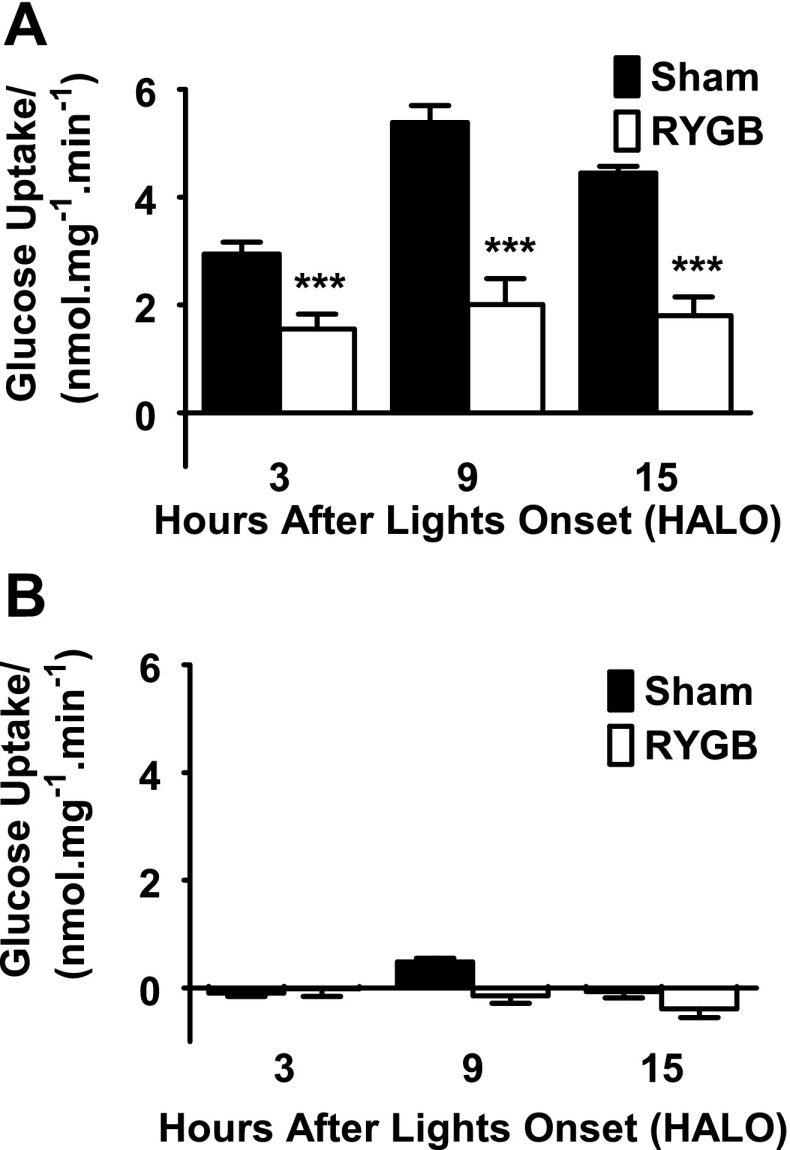
Fig. 5.
A: intestinal glucose transport capacity by time. Glucose uptake capacity in sham jejunum showed a normal diurnal rhythm, peaking at hours after lights on (HALO)-9 just prior to onset of feeding. This was completely abolished after RYGB. Glucose uptake capacity was significantly reduced in RYGB animals at all 3 times (***P < 0.001 compared with shams). B: the assay repeated in the presence of 20 μm phloridzin, confirming measured glucose transport represents Sglt1.

