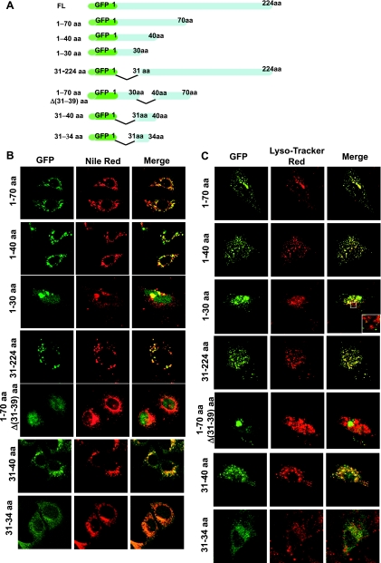
Fig. 3.
Expression of GFP-tagged Prdx6 deletion mutants in MLE-12 and A549 lung epithelial cells. A: schematic representation of GFP:Prdx6 deletion constructs. B: colocalization of each of the GFP:Prdx6 deletion mutants with Nile Red in MLE-12 cells. C: colocalization of Prdx6 with LysoTracker Red in A549 cells. Left panels: fusion constructs expressed in transfected cells. Middle panels: lamellar bodies (B) and lysosomes (C), shown in red. Right panels: colocalization, detected by yellow color, in the merged image. Inset in C demonstrates the absence of colocalization of GFP:Prdx6 (1–30 aa) in lysosomes.

