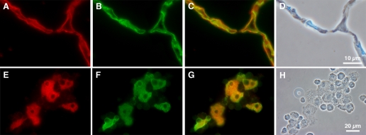
Fig. 10.
Immunohistochemical evidence of CLC5 in rat alveolar type I cells. Two-micrometer sections of adult rat lung (A–D) and cytocentrifuged preparations of adult rat mixed lung cells (E–H) were double stained with an antibody to CLC5 (red) and RTI40, an apical plasma membrane marker for TI cells (green). A: tissue section stained with CLC5 (red); B: same tissue section stained with RTI40 to display TI cells; C: merged image demonstrating colocalization of CLC5 and RTI40; D: phase-contrast image; of note, there are no TII cells in this section of tissue. E: mixed lung cell preparation stained with CLC5; F: cells stained with RTI40, which appear to be the same cells that stain positive for CLC5; G: merged image confirming colocalized signal; H: phase-contrast image of the cells; noteworthy are TII cells in the phase image are devoid of CLC staining in E. Results indicate the presence of CLC5 in TI cells.

