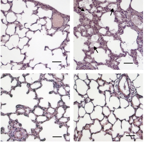
Fig. 5.
Formation of alveolar edema in HV animals (top, right) compared with normal controls (top, left) is blocked by EphA2/Fc (bottom, left) or anti-EphA2 antibody (bottom, right), shown by hematoxylin and eosin staining of microwave-fixed lung tissue. Note proteinaceous flooding of alveoli in hypoxic infected lung (arrows). Scale bar = 50 μm.

