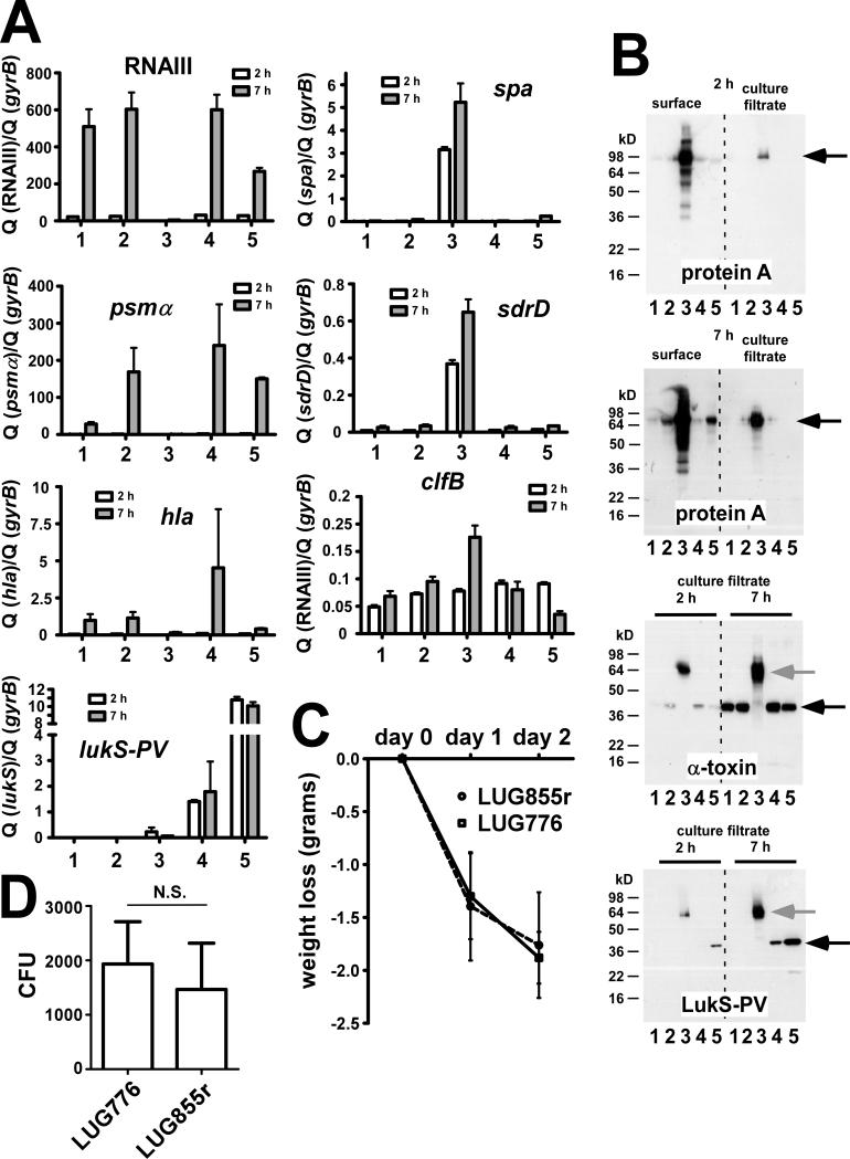
Figure 3.
Gene and protein expression of various key virulence factors and mouse pneumonia model. (A), Transcription levels by qRT-PCR. lukS-PV: lukS component of PVL. (B), Immunoblots of protein A, α-toxin, and the LukS component of PVL, using specific antisera (protein A, α-toxin: commercially available from Sigma; LukS-PV: produced by GenScript). Culture filtrate samples were directly used for SDS-PAGE; surface protein samples were obtained by lysostaphin digestion. Black arrow, detected protein, grey arrow, protein A reaction with IgG Fc part. (A,B) 1, RN6390; 2, LUG776; 3, LUG855 (original); 4, LUG855 (repaired); 5, LUG862. (C,D) Animal model of murine pneumonia with strains LUG776 and the isogenic, repaired LUG855r. The model was performed as described by Labandeira-Rey et al. [9], using the same mouse strain, inocula, and experimental conditions. Fifteen mice were used for each strain. Data for weight loss (C) and CFU/g in lung tissue samples (D) are shown. Statistical analysis was by unpaired Student's t-test. Error bars depict SEM.

