
Fig 2.
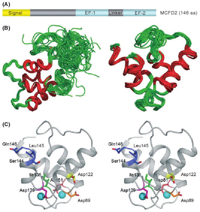
Structure of MCFD2 and locations of missense mutations. (A) MCFD2 primary sequence and domain structure. (B) The NMR structure of MCFD2 showing the complete structure (left) and only the ordered EF-hand domains (right). Red lines represent the alpha helices of the EF-hands. (C) A stereo view showing the positions of MCFD2 missense and C-terminal deletion mutations in the ordered structure of wild-type EF-hand domains (shown in colour). The two spheres represent calcium ions. Adapted from Guy et al (2008), with permission from Elsevier.