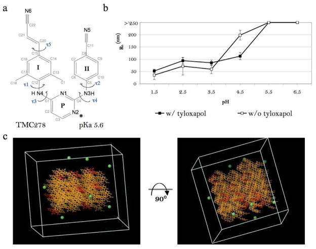
Figure 1.
Surfactant-dependent drug aggregation in vitro and the 10% protonated aggregate model in silico. a. Molecular structure of 1 with wing I, wing II, and pyrimidine rings labeled as I, II, and P respectively. (*) denotes the preferred protonation site at the N(2) atom of the pyrimidine ring, as suggested by the small-molecule crystal structure of protonated compound 145. b. Hydrodynamic radius distribution as a function of solution pH for 0.1 mM 1 in the presence and the absence of 0.1% tyloxapol. c. The starting conformation of the 10% protonated model in a 60 × 60 × 60 Å cell. Color assignment: yellow – neutral compound 1; red – charged compound 1; green – chloride ion. SPC water molecules are not shown.

