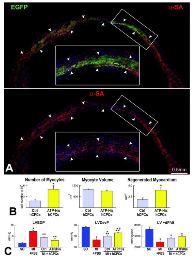
Figure 8.

Myocardial regeneration by activated hCPC. A, Mouse heart treated with histamine-stimulated hCPCs. The mid-portion of the infarct is replaced by EGFP-positive (upper panel, green) α-SA-positive cardiomyocytes (lower panel, red). The area in the rectangle is shown at higher magnification in the inset. B, Extent of regeneration mediated by hCPCs non activated (Ctrl hCPCs) or exposed to ATP or histamine (ATP-His hCPCs). C, LV function in sham operated (SO), infarcted untreated (MI + PBS) and hCPC-treated (MI + hSCPs) mice 7 days after coronary ligation. Ctrl, ATP and His identify non-stimulated, ATP-stimulated and histamine-stimulated hCPCs, respectively. LVEDP, LV end-diastolic pressure; LVDevP and +dP/dt. *P<0.05 vs. SO, **P<0.05 vs. MI + PBS, † P<0.05 vs. MI injected with untreated hCPCs.
