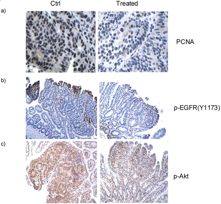
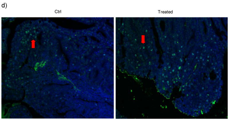
Figure 5. Immunohistochemical staining showing the in vivo effect of Sb on EGFR and Akt phosphorylation and on cell proliferation in the intestinal tumors of ApcMin mice.


a) The murine small intestines were stained using PCNA antibodies and counter-stained with haematoxylin. Dark/black colored nuclear staining indicates PCNA positive cells. Cells with blue colored nuclei are PCNA negative. Semi-quantative analysis using 5 representative images from each group showed that tumors from ApcMin mice treated with Sb possess fewer proliferative cells compared to those from control mice (37 ± 20 % vs. 66 ± 14%, p<0.01).
b&c) Antibodies against Phospho-EGFR(Y1173) and Phospho-Akt were used to stain the intestinal tumors counter-stained with haemotoxylin. From microscopic views of 10 tumors from each group, the percentage of phospho-EGFR (Tyr1173) positive cells per high power field in Sb-treated mice vs. control mice are 6.5 ± 2.8 % vs. 17 ± 5.5 %, (mean ± S.E., p<0.05). The average percentage of p-Akt strongly stained cells per high power field from Sb-treated mice vs. control mice are 49.6 ± 5.4% vs. 70.4 ± 4.5%, p=0.01. Representative images are shown to compare the different staining intensity for p-EGFR and p-Akt between treated and control ApcMin mice. (Brown color = Phospho-EGFR or Phospho-Akt).
d) Tunel staining of intestinal tissues from Sb-treated and control Apcmin mice. FITC (green) labeled cells are apoptotic tumor cells with DAPI (blue) staining as background. From 10 microscopic views each from the treated and control groups, the average percentages of apoptotic cells per high power field are 15 ± 3.0% in Sb-treated mice, vs. 4.5 ± 2.3 % in control mice respectively, (mean ± S.E., p<0.01). Representative tunnel staining images are shown Figure 5d with red arrow pointing to apoptotic cells within the tumors.
