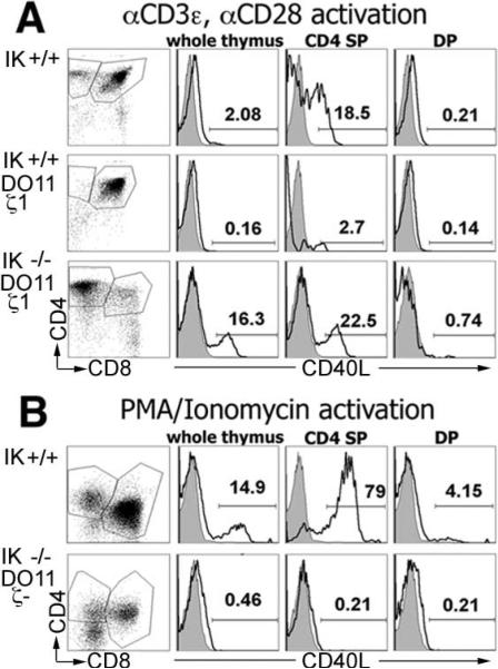
FIGURE 2.
CD4 SP cells that develop with reduced TCR signaling potential in the absence of IK are mature. Thymocytes were activated followed by staining with anti-CD4-allophycocyanin, anti-CD8α-FITC, and anti-CD40L-PE. Representative flow cytometry plots (far left column) of postactivation thymocytes with CD4 SP and DP gates that were used to analyze the up-regulation of CD40L are shown. Representative histograms (right columns) of anti-CD40L-PE staining (black line) compared with isotype control (shaded) in postactivation whole thymus, CD4 SP, and DP thymocytes are shown. A, Thymocytes from IK+/+,IK+/+ DO11.10 ζ1Tg, and IK–/– DO11.10 ζ1Tg mice activated with anti-CD3ε and anti-CD28. B, Thymocytes from IK+/+ and IK–/– DO11.10 ζ–/– mice activated with PMA/ionomycin.

