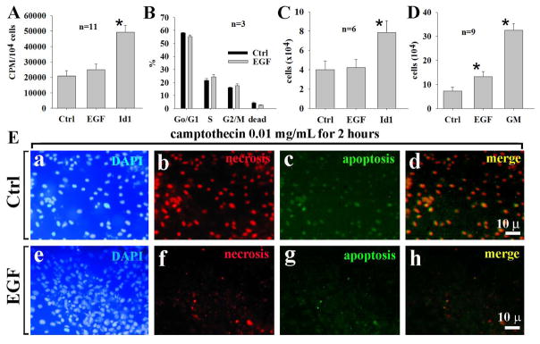
Fig. 2.
Commitment of EGF to cellular survival but not cell proliferation of OC1. In 6-hour incubation, EGF had no significant effects on the 3H-thymidine incorporation in OC1 cells compared with controls (A). Similarly, EGF had no effect on cell cycle progression from Go/G1-phase to S-phase within 6 hours compared with controls (B) and cell numbers (C). However, in 3-day cultures, EGF had more cell counts than control (D), due to an increase of cell survival. EGF significantly reduced cellular apoptosis and necrosis induced by camptothecin (E). Id1 served as a positive control for DNA synthesis (A) and cell counting (C). bar=10 μm applying to a-I; n=total samples from 3 separated experiments.

