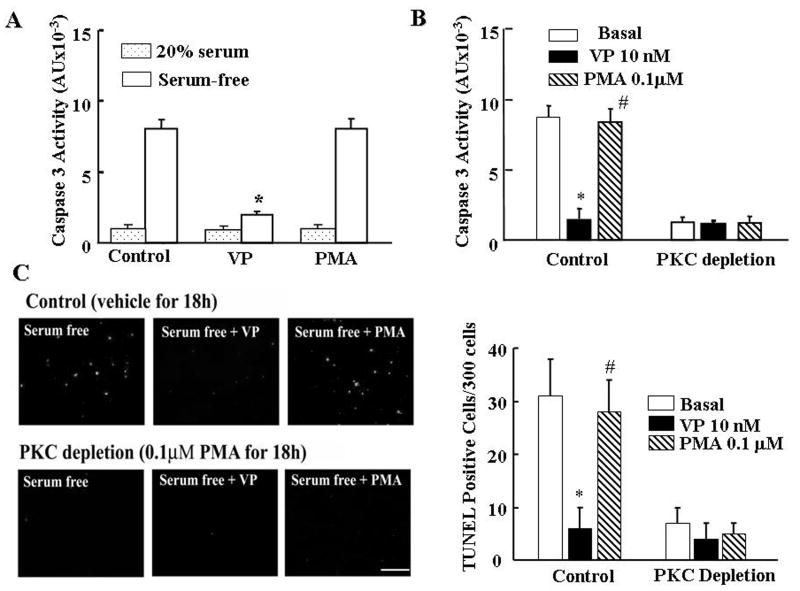
Fig.2.
The effect of stimulation PKC (acute-treatment with PMA) or PKC depletion (24h-PMA treatment) on serum-deprivation induced apoptosis in H32 cells. (A) Caspase 3 activity in H32 hypothalamic cells incubated in the presence of 20% serum or serum-free medium for 6h, in the presence or absence of 10nM VP or 100nM PMA. (B) Effect of PKC depletion by prolonged PMA treatment on caspase 3 activity in H32 cells. Following 24h exposure to PMA (100nM) cells were incubated in serum-free medium (Basal) in the presence or absence of 10nM VP or 100nM PMA for 6h. (C) TUNEL staining of control or PKC depleted H32 cells (PMA 100nM for 18h), following incubation for 6h in serum-free medium (Basal) in the presence or absence of 10nM VP or 100nM PMA. Scale bar, 100 μm. Bars represent the average ± S.E.M of the percent of TUNEL stained cells in a total of about 300 cells counted in 3 independent experiments conducted in duplicate. * p< 0.05, compared to Basal (serum-free) group; # p< 0.05 compared to serum-free + VP group.

