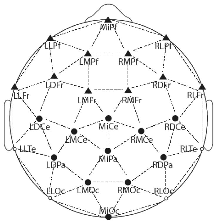
Figure 1.

Shown are the locations of the 26 scalp electrodes, as seen from the top of the head (with the front of the head at the top of the figure). Frontal electrodes are represented as triangles and central/posterior electrodes are represented as circles. The electrodes used for statistical analysis are shown using filled in shapes.
