
Figure 2.
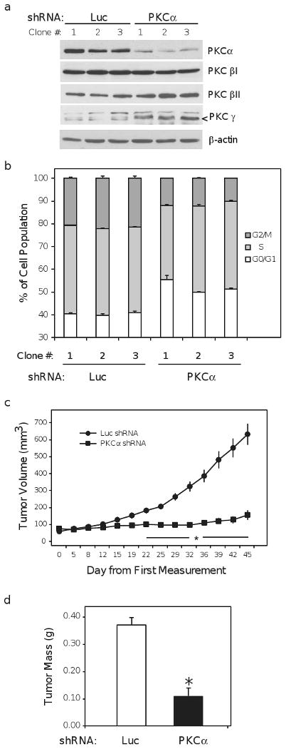
PKCα knockdown impairs tumorigenesis in vivo. (a) Ishikawa cell clones expressing luciferase (Luc) or PKCα shRNA constructs were isolated as described in materials and methods. Lysate from clonal populations was harvested after 24h of serum deprivation and probed for PKCα PKCβI, PKCβII, and PKCγ. β-actin is included as loading control and arrow (<) indicates the correct molecular weight band for PKCγ. (b) Luc and PKCα shRNA clones were deprived of serum for 24h and the proportion of cells in the indicated phases of the cell cycle was determined by flow cytometry. Data represent an average derived from two independent experiments. (c) Volume of xenograft tumors resulting from the subcutaneous injection of 10 million cells from Luc and PKCα shRNA clonal cell lines into athymic, nude mice. Each PKCα knockdown clone was injected contra-laterally to a Luc control in a total of 5 mice, thus data represent the combined average from n = 15 animals. (d) Average final weight of excised xenograft tumors assessed after the final dimension measurement. Bar graphs represent mean ± SEM; * denotes P < 0.01.