
Figure 6.
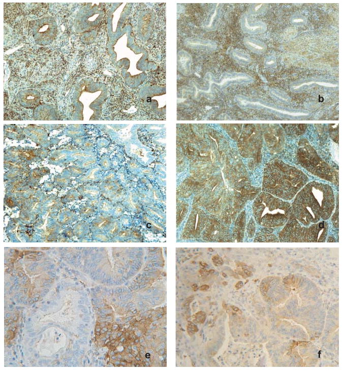
PKCα is abnormally expressed in endometrial tumors. Representative images of immunohistochemical staining for PKCα in normal proliferative (a) and secretory (b) endometrium. Staining in normal glandular epithelial cells was typically uniform and of low intensity; whereas endometrial stroma showed variable expression and was generally higher in stroma from secretory endometrium. Original magnification, ×100. (c) and (d), PKCα expression in representative grade 1 endometrioid adenocarcinomas. Original magnification, ×100. (c) typical PKCα staining in tumors was highly variable, with more intense staining frequently observed in discrete foci. (d) PKCα staining observed in a subset of tumors where high intensity expression occurred throughout the majority of the tissue section. (e) and (f), images from gland-like structures in endometrial tumors displaying focal variation in PKCα staining. Original magnification, ×250 and ×160, respectively.