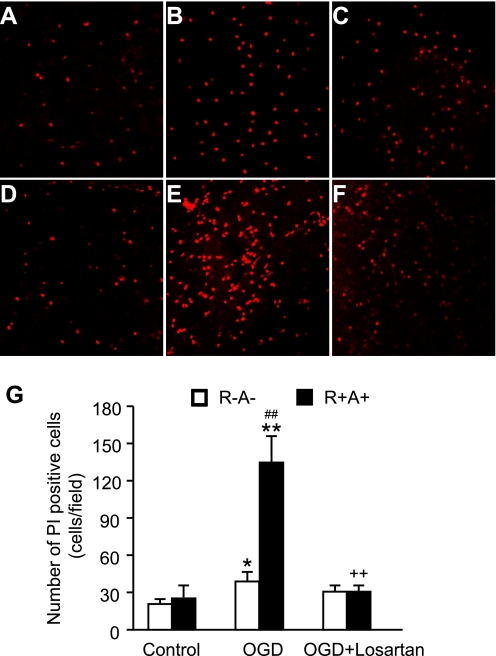
Fig. 4.
Exaggerated cerebral cortex cell death in R+A+ mice after OGD and the effect of losartan. Top: representative laser confocal microscopic images of propidium iodide (PI)-labeled cells in R−A− mice (A: OGD control, B: OGD; C: OGD + losartan) and R+A+ mice (D: OGD control, E: OGD; F: OGD + losartan). G: summarized data in PI-labeled cells after OGD. Data are expressed as means ± SE. *P < 0.05, **P < 0.01, OGD vs. OGD control; ++P < 0.01, OGD + losartan vs. OGD; ##P < 0.01, R+A+ vs. R−A−.

