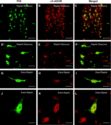
Fig. 3.
Colocalization of α4-nAChRs and 5-HT neurons in the fetal baboon medulla. A, D, G, and J: PH8, a marker for tryptophan hydroxylase, the key enzyme in the synthesis of 5-HT, immunopositive neurons (green). B, E, H, and K: α4 immunopositive cells (red) in the raphé obscurus (A–F) and extra-raphé (paragigantocellularis lateralis) (G–L) at 161 days of gestation (term = 180 days). C, F, I, and L: colocalization of expression is demonstrated by merging of images (yellow). A subpopulation of neurons expressing α4, which were not 5-HT, were observed in association to 5-HT cells. Scales: A–C: 115 μm; D–F: 57 μm; G–I: 30 μm; J–L: 35 μm.

