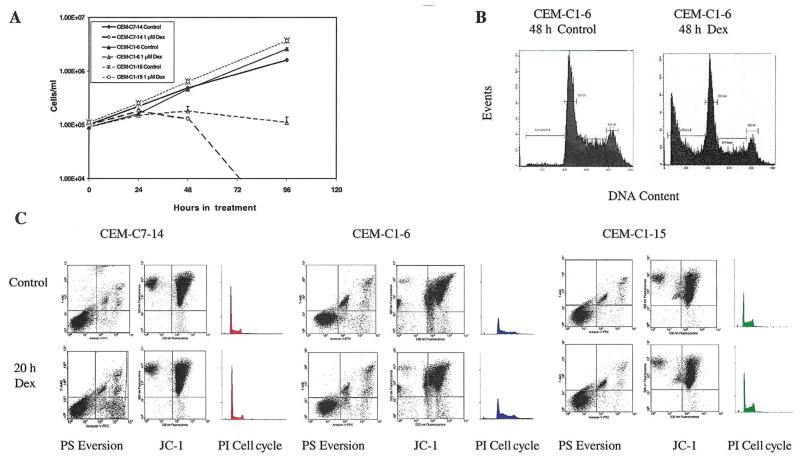
Fig. 1.
Characteristics of CEM clones. (A) Growth curves for CEM-C7-14, CEM-C1-6, and CEM-C1-15 cells treated with vehicle (Control) or 1 μM Dex. Cell viability was determined using Trypan Blue exclusion and a hemacytometer. (B) Propidium iodide (PI) analysis of CEM-C1-6 cells for apoptotic, sub-G1 levels of DNA after 48 h of vehicle (Control) or 1 μM Dex treatment. (C) Flow-cytometric analyses of CEM-C7-14, CEM- C1-6, and CEM-C1-15 cells treated with vehicle (Control) or 1 μM Dex for 20 h. Cells were evaluated using Annexin-V-FTC/7AA-D to assess phosphatidylserine (PS) membrane eversion (lower right quadrant = intact cells with everted PS), JC-1 to assess mitochondrial integrity (lower right quadrant = depolarized mitochondria), and PI staining of DNA for apoptosis and cell cycle analysis (subdiploid apoptotic population left of initial peak).

