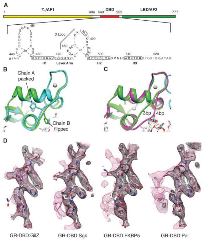
Fig. 2.
DNA sequence-mediated structural differences in GR-DBD. (A) Domain structure of GR. τ1, tau1. (B) Overlay of chains A and B from GR-DBD:Pal complex shows packed and flipped conformations. (C) Overlay of chain B from GR-DBD complexed with 4-bp spacer (15) (magenta) and 3-bp spacer GBS (green). (D) Composite omit maps of GR-DBD complexed with different GBSs (GilZ, FKBP5, Sgk, and Pal) under the same conditions. Lever arm peptide is shown with 2Fo-Fc (black mesh) and composite omit map (red mesh) overlaid.

