
Figure 2.
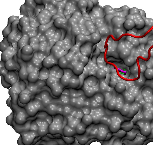
Example of a lysine hot spot. A detail of the interaction between trypsin (in grey, surface representation) and trypsin inhibitor (in red) is shown (pdb code 2PTC). The side chain of the hot spot Lys15 from the inhibitor (in magenta, ΔΔG = 10 kcal/mol) fits into a hole on the trypsin surface. The ζ-nitrogen of the Lys side chain forms two hydrogen bonds (highlighted in green) with the residue Ser190 from trypsin.