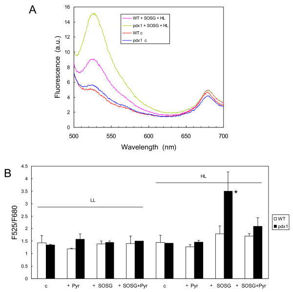
Figure 7.
Fluorescence of SOGS in WT and mutant (pdx1) leaves exposed to high light. A) Fluorescence of leaves infiltrated with SOGS after exposure to white light (HL = 450 μmol photon m-2 s-1 for 40 min). Controls (= c) were kept in dim light before fluorescence measurements. B) Fluorescence ratio F525/F680 of WT leaves and mutant leaves infiltrated with SOGS and/or vitamin B6 before or after illumination. Data are mean values of 3 measurements + SD. *, significantly different from the WT value with P < 0.025 (t test).

