Figure 2.
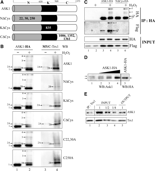
Identification of the cysteines in ASK1 responsible for the covalent association with Trx1. (A) Schematic representation of the different cysteine substitution mutants used. Represented are the N-terminal (N), kinase (K), and C-terminal (C) domain of ASK1. Residue numbers at domain boundaries are indicated. Numbers in bold or in white within each domain indicate which cysteine residue(s) have been replaced by alanine residue(s). (B and C) 293T cells were transfected with pRK-Flag-Trx1 alone (C), or pRK-Flag-Trx1 or pCMV5-Myc-Trx1 together with the various ASK1 constructs defined in A or other labeled “Cx,yA” (the x and y indicates the positions of ASK1 cysteines residues which have been replaced by alanine residues) as indicated at the right (B) or the top (C) of panels. Twenty-four hours after transfection, the cells were left untreated (−) or exposed (+) to 1 mM H2O2 for 3 min. Total cell extracts were migrated on nonreducing (B) or reducing (INPUT in C) SDS gels, and anti-HA immunoprecipitated extracts were migrated under nonreducing conditions (IP:HA in C). Western blot (WB) analysis were performed either with anti-HA (ASK1-HA in B and HA in C), anti-MYC (MYC-Trx1 in B), or anti-Flag (Flag in C). Band 1 indicates the expected positions of monomeric ASK1 (150 kDa), bands 3 and 4 point the position of ASK1 disulfide-bonded complexes, and bands 2A and 2B indicate the positions of Trx1-ASK1 covalent complexes. (D) HEK293 cell extract (− was combined with increasing amounts of extracts from HEK293 cells transfected with pcDNA3-ASK1-HA. Total μg of proteins was equivalent between each lane. Extracts were migrated on SDS gels under nonreducing conditions and analyzed by Western blot with either anti-ASK1 (WB:ASK1) or anti-HA (WB:HA). The pcDNA3-ASK1-HA extracts in lanes 4 and 5 are the same. Arrows indicate the expected positions of monomeric endogenous ASK1. (E) 293T cells extracts were immunoprecipitated with anti-Trx1. Total cell (INPUT), anti-Trx1 immunoprecipitated (IP:Trx1), and anti-Trx1 depleted (OUTPUT) extracts were migrated on reducing SDS gels and analyzed with either anti-ASK1 (ASK1) or anti-Trx1 (Trx1). The third (labeled “1/2”) and fourth (labeled “1/4”) lanes were loaded with two and four times less protein than the second lane. They serve as a scale to determine the quantity of Trx1 and ASK1 depleted from the INPUT when compared with the OUTPUT. Lane numbers are referred to in the text.

