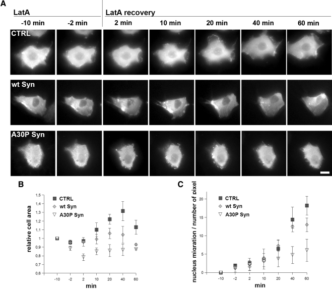
Figure 7.
Time-lapse microscopy of MDCK cells during LatA treatment and washout. A30P α-synuclein expression inhibits actin reorganization, lamellipodia formation and cell movement. (A) The three rows show frames from time-lapse microscopy videos (Videos 1–3) of CTRL, wt α-synuclein, or A30P α-synuclein–expressing MDCK cell transfected with actin-GFP, during LatA treatment and washout. Notice in the top row the appearance of lamellipodia guiding the cell movement during wash out of LatA; in the low bottom row the appearance, already at 2 min, of large subplasmalemma GFP-actin aggregates persisting up to the end of the experiment. (B) Relative changes of the average cells area in MDCK cells during LatA incubation and recovery as in A. The area is normalized to the −10-min time point. (C) Changes of the rate of cell movement in MDCK during LatA incubation and recovery as in A, expressed as the traveled distance of the nucleus. The points in B and C are averages (±SEM) of three different measurements from independent experiments. Bar, 10 μm.

