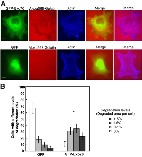
Figure 2.
Overexpression of Exo70 stimulates invadopodial activity. (A) MDA-MB-231 cells were transfected with GFP-tagged Exo70 (GFP-Exo70), plated on fluorescent Alexa 568–labeled gelatin film for 4 h, and then processed for microscopy. Individual and merged images of GFP-Exo70 fluorescence (green), F-actin (blue), and gelatin (red) are shown. Overexpression of GFP-Exo70 stimulated focal degradation in MDA-MB-231 cells (top panel). Cells transfected with GFP alone were used as negative control (bottom panel). (B) Quantification of degradation areas in cells overexpressing Exo70. Three independent measurements (∼70 cells each) for each treatment were carried out. Error bars, SD. *p < 0.01. Scale bar, 5 μm.

