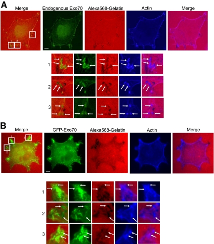
Figure 3.
Localization of Exo70 at the focal degradation sites. (A) MDA-MB-231 cells were plated on fluorescent Alexa 568–labeled gelatin (red) for 20 h. After fixation, cells were stained for Exo70 (green). Individual and merged images are shown. Endogenous Exo70 showed colocalization with the “degradation holes” in the fluorescent gelatin matrix, along with some enrichment at the plasma membrane. (B) The localization of GFP-tagged Exo70 expressed at low levels using the pJ3-GFP vector was examined in MDA-MB-231 cells. GFP-tagged Exo70 partially overlapped with the degrading spots. Higher magnification views of the boxed areas are shown underneath each image. Arrows, colocalization of Exo70 and invadopodia-forming sites. Scale bars, 5 μm.

