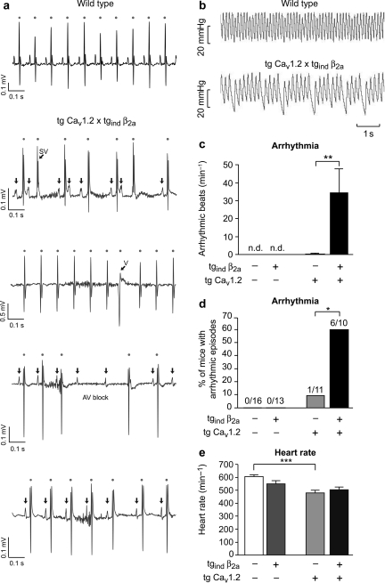
Figure 4.
Arrhythmia in transgenic mice expressing β2a and CaV1.2 Ca2+-channel subunits. (A and B) Original traces of recordings obtained by ECG telemetry in awake, freely moving mice (A) from wild-type (upper panel) and double-transgenic mice (lower panels) or by microtip catheterization in anaesthetized animals (B), respectively. (A) In case of wild-type mice (top row) we observed sinus rhythm as displayed. Tg CaV1.2 × tgind β2a mice showed several forms of rhythm disturbances including supraventricular extrasystolies (second row, SV), ventricular extrasystolies (third row, V), atrio-ventricular block (fourth row), and sinus pause/sinus bradycardia (bottom row). (B) Catheter data revealed the haemodynamic relevance of rhythm disturbances in double-transgenic mice. (C) The incidence of arrhythmic beats during microtip catheterization was significantly increased in tg CaV1.2 × tgind β2a mice (**P < 0.01). No arrhythmic beats were detected in wild-type or tgindβ2a mice. (D) Overall, arrhythmic episodes during ECG or catheterization were only seen in 1/11 (9.1%) tg CaV1.2, but in 6/10 (60%) double-transgenic mice (*P < 0.05; χ2 test). (E) Heart rate as determined by ECG telemetry in awake animals was significantly lowered in tg CaV1.2 mice (n = 5–9 per genotype, 3/5 examined tg CaV1.2 mice did not receive tebufenozide, *P < 0.05, **P < 0.01; ***P < 0.001).

