Figure 6.
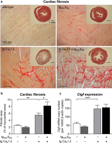
Cardiac fibrosis in transgenic mice overexpressing CaV1.2 Ca2+-channel subunits. (A) Sirius-red staining of mid-ventricular cardiac sections from wild-type mice and mice overexpressing the pore-forming subunit CaV1.2, the auxiliary subunit β2a or both subunits, respectively. Hearts of mice overexpressing CaV1.2 or additionally β2a (tg CaV1.2 × tgind β2a) displayed marked fibrosis. Inserts show heart slices stained with haematoxylin–eosin. (B) Interstitial fibrosis was determined by Sirius-red staining and is displayed as percentage of left ventricular cross-sectional area. Overexpression of CaV1.2 lead to significant increases in interstitial fibrous tissue (n = 8–16 per genotype, *P < 0.05, **P < 0.01). (C) mRNA expression of connective tissue growth factor (Ctgf) was quantified by qPCR. In cardiac ventricles of tg CaV1.2 and tg CaV1.2 × tgind β2a mice Ctgf expression was significantly increased (n = 8–15 per genotype, ***P < 0.001).

