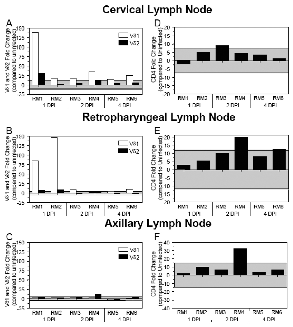
Figure 2. Increased Vδ1 and Vδ2 γδ TCR mRNA expression detected in the cervical and retropharyngeal LNs during acute SIV infection.
The fold change in Vδ1, Vδ2, and CD4 gene expression was assessed in the cervical (A and D), retropharyngeal (B and E), and axillary (C and F) lymph nodes of SIV+ rhesus macaques necropsied at the designated days following oral SIV inoculation. Changes in Vδ1 TCR expression are in white bars while changes in Vδ2 TCR expression are in black bars. The mRNA levels shown are reported as fold change with regard to the average of mRNA levels in matched samples of four uninfected macaques. The grey shaded area represents a two standard deviation range of the average expression in uninfected macaques. Bars extending beyond the grey shaded area represent samples that are increased or decreased with regard to the uninfected controls.

