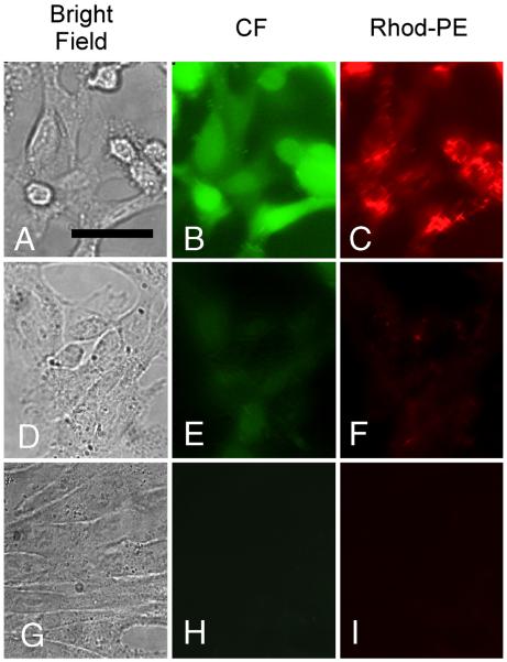
Figure 5. Fluorescence images of CV1 cells incubated with Rhod-PE–containing liposomes encapsulating CF.
Panels A, D and G are brightfield images; panels B, E and H are images acquired in the CF fluorescence channel; panels C, F and I are images acquired in the Rhod-PE channel. Scale bar in panel A represents 20 μm. Cells avidly endocytosed PS-bearing (negatively charged) liposomes at 37 °C, leading to bright intracellular CF and Rhod-PE signals (A-C). In contrast, neutral liposomes (bearing no PS) are poorly endocytosed at 37 °C (D–F). Endocytosis of negatively charged liposomes is blocked at 4° C, resulting in no observable uptake of CF or Rhod-PE (G–I).

