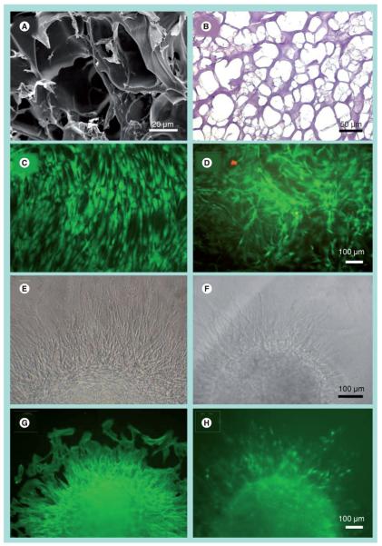
Figure 1. In vitro characterization of bone marrow-derived mesenchymal stem cell morphology and survival in 3D fibrin after 2 days.
Visualization of porous fibrin structure by (A) scanning electron microscopy and (B) hematoxylin and eosin of a cross-sectional slice. Live/Dead® viability assay depicts viable (green) and dead (red) cells on (C) control tissue culture dishes or in (D) fibrin gels. Migration of bone marrow-derived mesenchymal stem cells from aggregates on (E & G) control or (F & H) fibrin gels as shown by bright field (E & F) or by immunofluorescence staining of F-actin (G & H).

