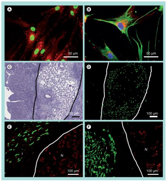
Figure 2. In vivo delivery of bone marrow-derived mesenchymal stem cells in fibrin into the infarcted myocardium.

Immunofluorescence staining of (A) human-specific nuclear matrix antigen (NuMA, green) and connexin-43 (red) and (B) human-specific vimentin (green), connexin-43 (red) and Hoechst nuclear dye (blue). At 2 days postinjection, fibrin was visible by (C) hematoxylin and eosin staining, and bone marrow-derived mesenchymal stem cells (BM-MSCs) were visualized by (D) NuMA-positive BM-MSCs in an adjacent tissue section. The boundary of the fibrin matrix is marked for clarity. BM-MSCs persisted in the infarct after 5 weeks of injection, as shown by immunofluorescence staining of (E) NuMA (green) and connexin-43 (red), and (F) vimentin (green) and connexin-43 (red). The boundaries between infarct and normal myocardium are marked for clarity.
I: Infarct; N: Normal.
