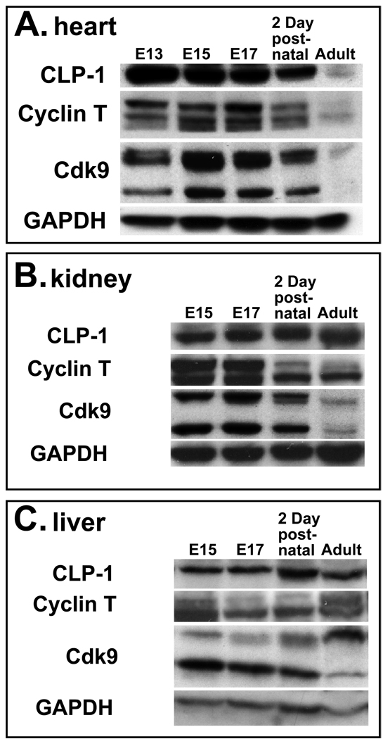
Figure 1.
Expression of CLP-1, cyclin T and cdk9 proteins in developing and post-natal heart (A), kidney (B), and liver (C). One hundred micrograms of protein lysate was loaded per lane and expression of CLP-1, cyclin T, cdk9, and GAPDH proteins detected by immunoblot analysis. GAPDH expression served as loading control. The E15 to E17 pre-natal period encompasses the time period in which CLP-1 knockout fetuses develop hypertrophy and die (24). Definitive liver and kidney tissues were not identifiable in E13 embryos and thus not harvested.

