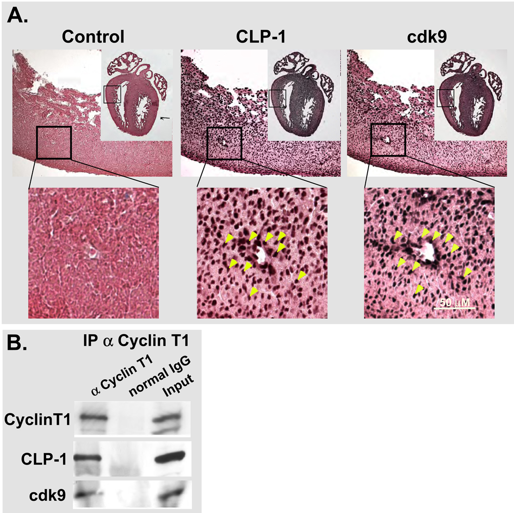
Figure 2.
Expression of cdk9 and CLP-1 in E17 and E19 heart. A. Serial paraffin sections from E17 mouse heart showing co-localization of cdk9 and CLP-1 in heart cells. Boxed regions of hearts show area depicted in images (a transected vein (white area in image) was used to co-align images for comparing overlap in nuclear stainings). B. Immunoprecipitation of E19 heart lysates showing co-immunoprecipitation of CLP-1 with P-TEFb components. Input refers to the non-precipitated lysate.

