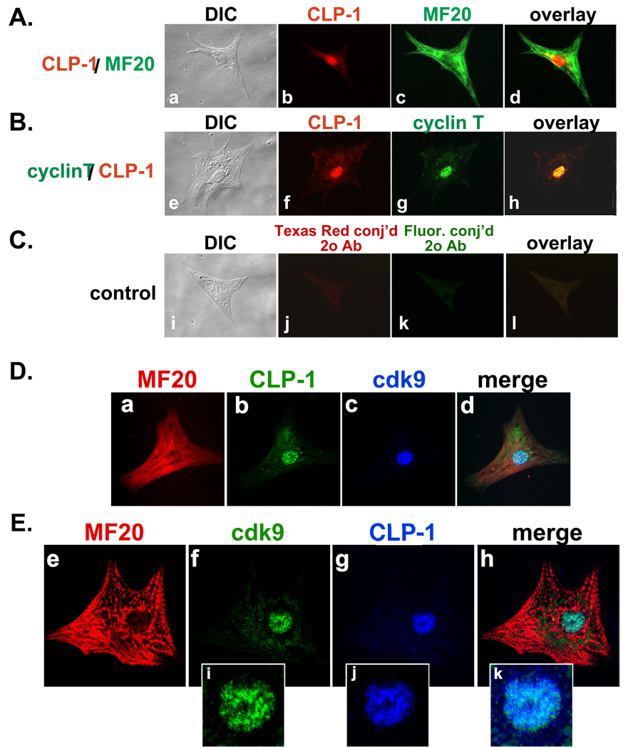
Figure 3.
Immunocytochemical co-localization of CLP-1, cyclin T and cdk9 in isolated 2 day post natal rat cardiomyocytes. A–C: (A) CLP-1 is in the nucleus of MF20-positive cardiomyocytes; (B) nuclear co-localization of CLP-1 and cyclin T; (C) control (no 1° antibody). (DIC, Differential Interference Contrast microscopy). D,E: Confocal microscopy of cardiomyocytes. D and E: CLP-1 and cdk9 are co-localized to the nucleus of MF20-positive cardiomyocytes. Panels i-k, magnified images of nuclei showing speckled type nuclear staining of cdk9 and CLP-1.

