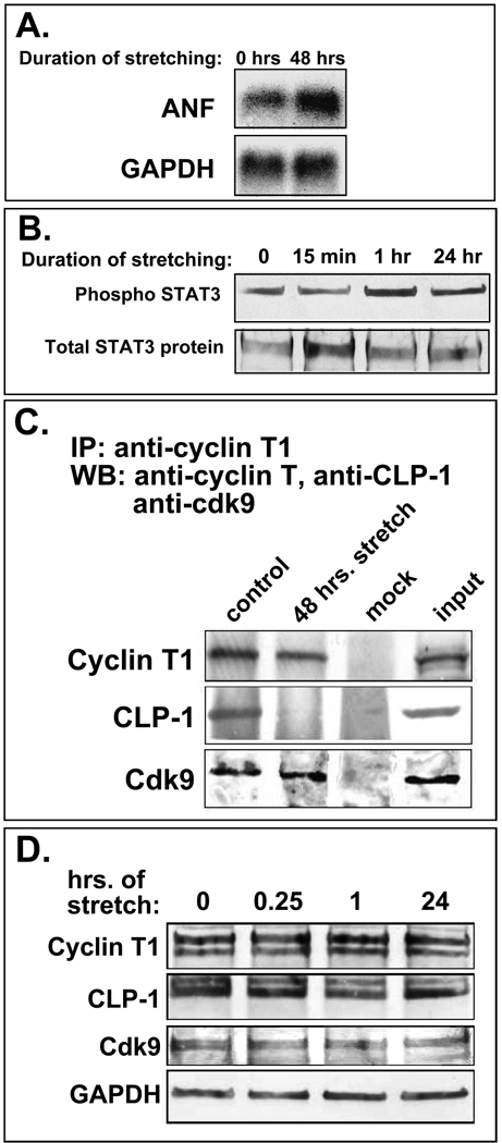
Figure 4.
A. Stretched cardiomyocytes express markers of hypertrophy. Northern blot showing increased ANF (atrial natriuretic factor) mRNA levels in stretched cardiomyocytes. GAPDH is loading control. B. Phosphorylation of STAT 3 increases with continued stretch of cardiomyocytes (1 hr and 24 hr); overall STAT3 protein levels remain constant. C. CLP-1 is released from p-TEFb complexes in stretch-induced hypertrophic cardiomyocytes. Immunoblot of p-TEFb components in stretched and non-stretched cardiomyocytes showing markedly reduced levels of CLP-1 in P-TEFb complexes from cardiomyocytes stretched for 48 hours; cyclin T1 and cdk9 levels remain unchanged. D. Total levels of p-TEFb components (free and complexed) remain unchanged with stretch. GAPDH is loading control.

