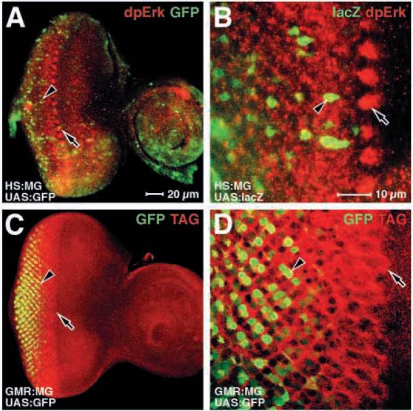
Fig. 3.
Neither dpErk antigen or the MG fusion protein are detected in cell nuclei in the morphogenetic furrow. Eye discs are shown; anterior rightwards. A,C and B,D are pairs at the same magnification. Genotypes are indicated in the bottom left-hand corner; antigens are indicated in the top right-hand corner. (A,B) The activity of the MG fusion protein in activating reporter genes (UAS:GFP and UAS:lacZ as indicated), relative to the endogenous dpErk antigen. Note that not only is reporter gene activity later than the high level dpErk antigen cell clusters in the furrow, but that the pattern of clusters is not reiterated by the reporter gene. (C,D) The same specimen showing the MG fusion protein expressed specifically behind the furrow (GMR:MG). Note that although the MG protein can be detected directly in most or all cells from the furrow (the leading edge of the TAG, arrow), MG activity is detected later and in a more restricted subset of cells (GFP, arrowhead).

