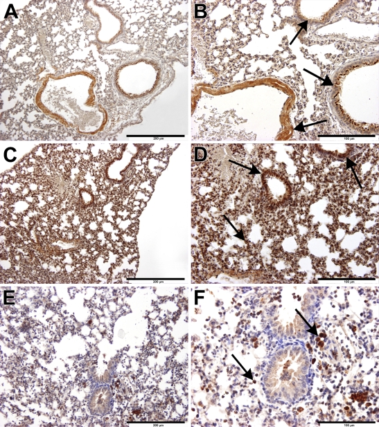
Figure 2.
Immunohistochemical localization of sst4 receptors in the mouse lung. Arrows indicate sst4 receptor positivity on epithelial/endothelial and smooth muscle cells, as well as on activated macrophages. (A,B) In the non-inflamed lung, sst4 immunopositivity is observed in bronchial epithelial cells, particularly on the luminal surfaces, vascular endothelial cells, and vascular and bronchial smooth-muscle cells in the interalveolar septal regions. (C,D) In the inflamed lung, 24 hr after intranasal LPS administration, there is a marked accumulation of sst4-immunopositive mononuclear cells, predominantly macrophages but also neutrophils and lymphocytes, in the peribronchial/perivascular spaces. (E,F) Three months of whole-body cigarette smoke exposure induces infiltration of sst4 receptor–expressing activated macrophages in the intraalveolar regions; there are also lymphocytes and a few granulocytes. Bars: A,C,E = 200 μm; B,D,F = 100 μm.

