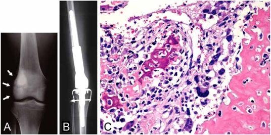
Fig.(1).
Osteoblastic osteosarcoma. Plain radiograph of a 19-year-old male clearly shows an irregular osteoblastic feature in the medullary region of the distal femur with cortical irregularity over the lateral cortex (arrows) (A). After a three-drug chemotherapy regimen made up of cispatin, doxorubicin and methotrexate, wide resection was carried out, followed by reconstruction using a Kotz prosthesis (HMRS: Howmedica Modular Reconstruction System) (B). Histological section reveals osteoblastic osteosarcoma clearly demonstrating osteoblastic features, comprising irregular immature bone deposition by anaplastic tumor cells (C).

