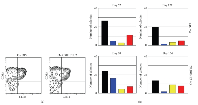
Figure 3.
Characterization of cultured cells. (a) Flow cytometric analysis of detached cells produced in cultures on OP9 (Exp-OP9-A) and C3H10T1/2 (Exp-10T1/2-A) feeder cells and collected on Day 218 of culture. The detached cells were stained for CD33, a marker specific for granulocyte/macrophage lineage cells, and CD34, a marker specific for hematopoietic stem/progenitor cells. Flow cytometric analyses of detached cells from other experiments showed similar results. (b) Colony-formation assays. Detached cells produced in culture on OP9 feeder cells (Exp-OP9-A) were collected on Days 57 and 127 of culture. Similarly, detached cells produced in culture on C3H10T1/2 feeder cells (Exp-10T1/2-D) were collected on Days 60 and 134 of culture. The cell samples were used in a standard colony-formation assay. Black bars: colony-forming unit of monocyte/macrophage lineage cells, CFU-M. Blue bars: colony-forming unit of granulocyte lineage cells, CFU-G. Yellow bars: colony-forming unit of granulocyte and monocyte/macrophage lineage cells, CFU-GM. Red bars: burst-forming unit of erythroid cells, BFU-E. Similar results were obtained in colony-formation assays using detached cells from other cultures.

