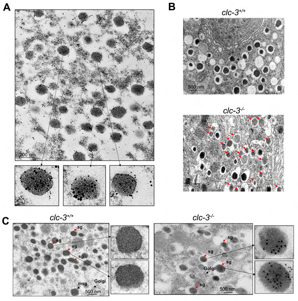
Figure 3. Electron microscopic localization of ClC-3 on insulin granules and characterization of secretory granules in thin sections of β–cells from clc-3+/+ and clc-3−/− mice.
(A) Immunogold staining shows insulin (10 nm particles) and ClC-3 (15 nm particles) colocalized in secretory granules of β–cells. (B) Comparison of clc-3+/+ and clc-3−/− cells reveals abundant pale, immature-appearing secretory granules in the knockout cells (marked by arrows). (C) Localization of proinsulin on thin sections of pancreatic β–cells from clc-3+/+ and clc-3−/− mice. In clc-3+/+ mice proinsulin staining is observed only in the nascent (maturing) secretory granules (msg). Mature secretory granules (sg) appear free of labeling. In the β–cells from the clc-3−/− mice, proinsulin labeling is found regardless of the state of maturity.

