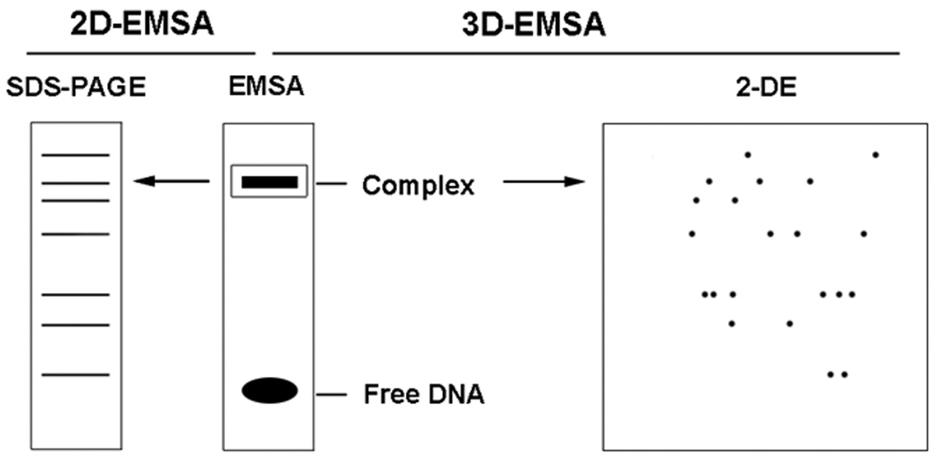
Fig.5.

Scheme of 2D-EMSA and 3D-EMSA. Nuclear extract is incubated with radiolabeled element DNA to form specific DNA-protein complex and analyzed by EMSA. The complex on the non-denature PAGE gel is cut out, crashed and mixed with 1X Laemmli buffer or Rehydration buffer for further separation by SDS-PAGE (2D-EMSA)or two-dimensional electrophoresis (3D-EMSA).
