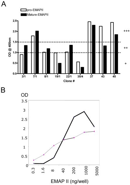
Figure 1. Analysis of anti-EMAP II antibodies by ELISA.
A) Monoclonal anti-EMAP II antibodies recognize full length and processed EMAP II in ELISA. Subclones of monoclonal anti-EMAP II antibodies were tested in ELISA using recombinant EMAP II protein and the response was graded as + (slightly positive; OD <1.0), ++ (medium positive; OD >1.0 & <1.5), +++ (strongly positive; OD >1.5) based on the optical densities. Data shown are from a representative experiments repeated two times independently with similar results. B) Dose dependence of EMAP II. Comparison of EMAP II polyclonal (gray line) and M7/1 monoclonal antibodies (bold line) by ELISA. Shown are the OD values from the ELISA obtained with increasing concentrations of EMAP II.

