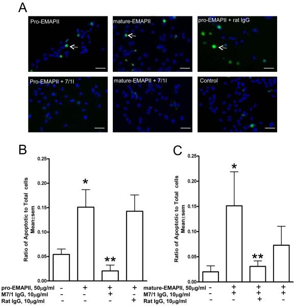
Figure 4. Inhibition of EMAP II-induced apoptosis in endothelial cells.
A) Endothelial cells incubated with pro-EMAPII protein (50 μg/ml) or mature-EMAPII protein (50 μg/ml) demonstrated a significant apoptosis (arrows) as shown by TUNEL (*p<0.01). Pre-treatment of these cells with neutralizing antibody M 7/1 (10 μg/ml) but not with control rat IgG significantly (**p<0.03) inhibited apoptosis induced by both pro and mature EMAPII as shown from representative fluorescent microscope images following TUNEL assay. B) and C) Quantification of TUNEL positive cells by MetaMorph software normalized to total DAPI nuclear positive cells is shown below for pro-EMAPII (B) and mature EMAPII (C). Data shown are from a representative experiment performed in triplicates and repeated independently two additional times with similar results. Scale bar=50μm

