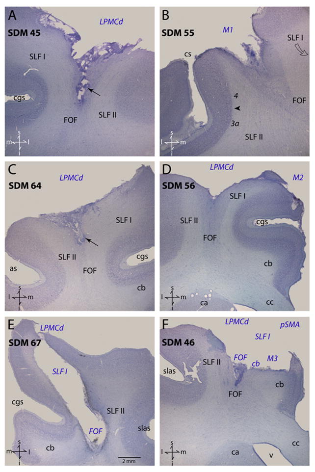
Figure 1.

Plate of photomicrographs illustrating Nissl-stained coronal sections through representative cases analyzed in this study. In each panel the region of extirpated cortex and involved subcortical pathways are identified by the blue italicized conventions. The arrows in panels A and C identify the tapered portion of the subcortical lesion that part the adjacent SLF I and SLF II. The arrow head in panel B identifies the boundary between Brodmann’s areas 4 and 3a. The scale bar in panel E corresponds to all panels. Directional orientation is indicated in the lower left corner of each panel with abbreviated directions: superior (s), inferior (i), medial (m) and lateral (l). Abbreviations: as, spur of the arcuate sulcus; ca, caudate nucleus; cb, cingulum bundle; cc, corpus callosum; cgs, cingulate sulcus; cs, central sulcus; FOF, fronto-occipital fasciculus; i, inferior; l, lateral; LPMCd, dorsal lateral premotor cortex; m, medial; M1, primary motor cortex; M2, supplementary motor cortex; pSMA, pre-supplementary motor cortex; s, superior; slas, superior limb of the arcuate sulcus; SLF I, superior longitudinal fasciculus I; SLF II, superior longitudinal fasciculus II; v ventricle.
