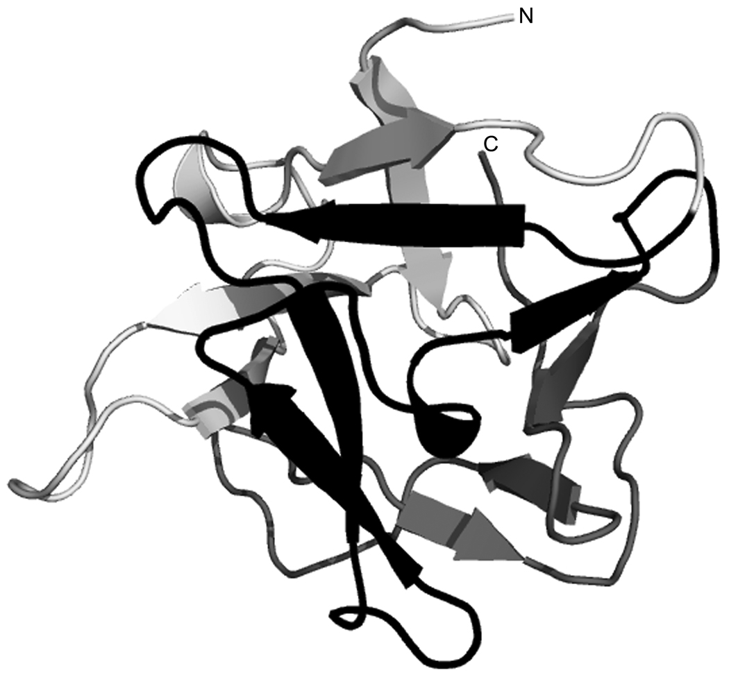
Fig. 3.
Ribbon diagram of the predicted three-dimensional structure of CNL. The lectin adopts a β-trefoil fold typical of ricin B-type lectins, consisting of three subdomains depicted in the following colors: α-repeat, light gray; β-repeat, black; γ-repeat, dark gray. N and C mark the N-and C-terminal ends of the domain.

