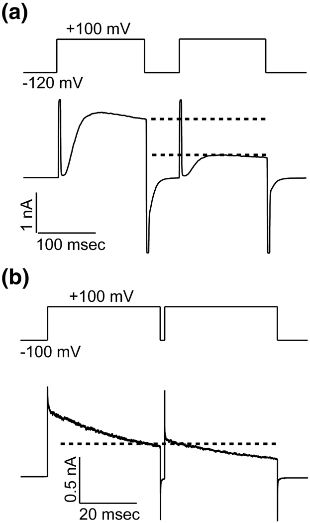
Figure 1. A Comparison of Inactivation in KvAP and Shaker Kv Channels.
DPhPC vesicles containing KvAP (A) or Kv1.2–Kv2.1 Paddle Chimaera (B) were fused into DPhPC bilayers. The current response to a paired depolarization pulse from a holding voltage −120 mV (KvAP, A) −100 mV (Paddle Chimaera, B) to +100 mV was recorded. The dotted lines indicated the current levels at the end of the first and the beginning of the second depolarization pulse.

