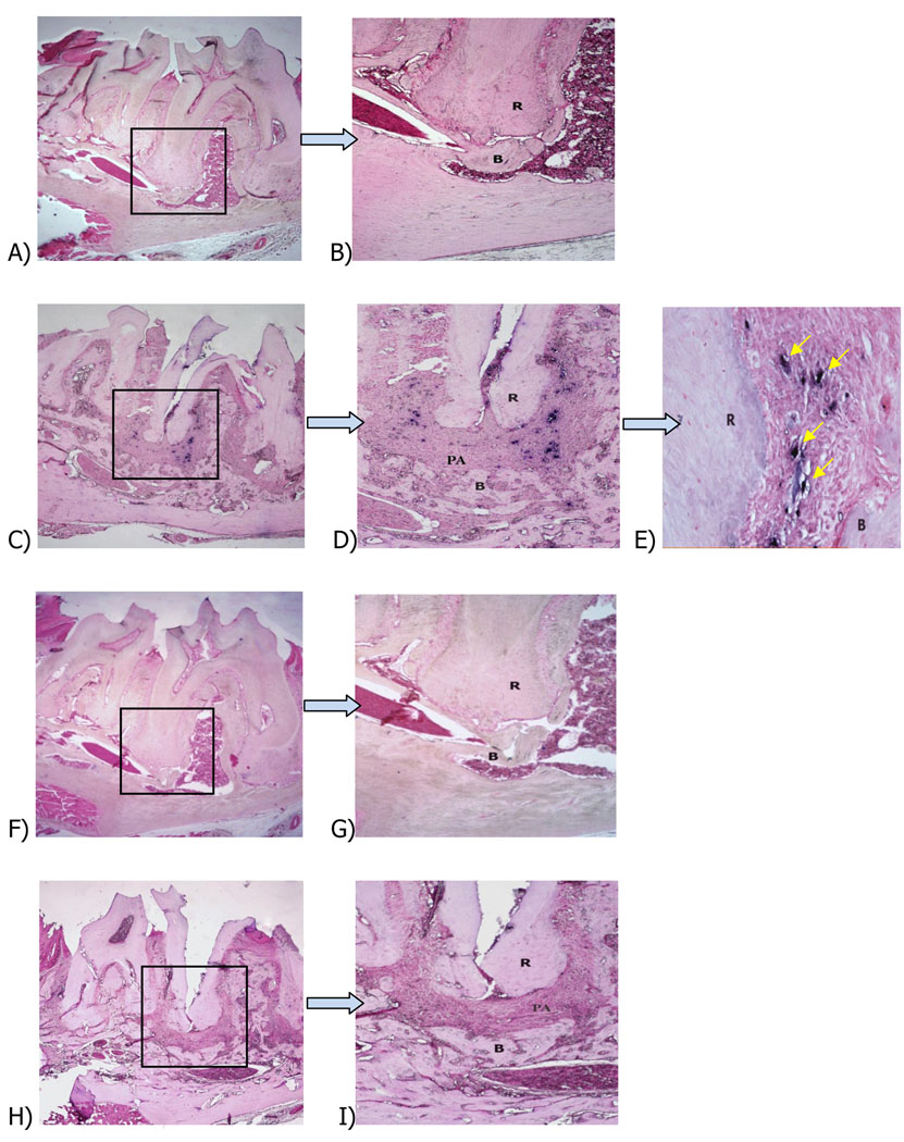
Figure 1. In situ hybridization of F0XP3+ cells in periapical lesions.
Representative sections of (A): F0XP3 anti-sense probe, control unexposed pulp, first mandibular molar (×40); (B): higher magnification (×l00 of box in A); (C): FOXP3 anti-sense probe, exposed pulp (×40); (D): higher magnification (×l00) from C; (E): higher magnification (×400) from D; (F): FOXP3 sense probe, control unexposed pulp; (G): higher magnification (×400) from F; (H): FOXP3 sense probe, exposed pulp; (I): higher magnification (×l00) from H; R: root; PA: periapical lesion; B: bone. Eosin counterstain.

