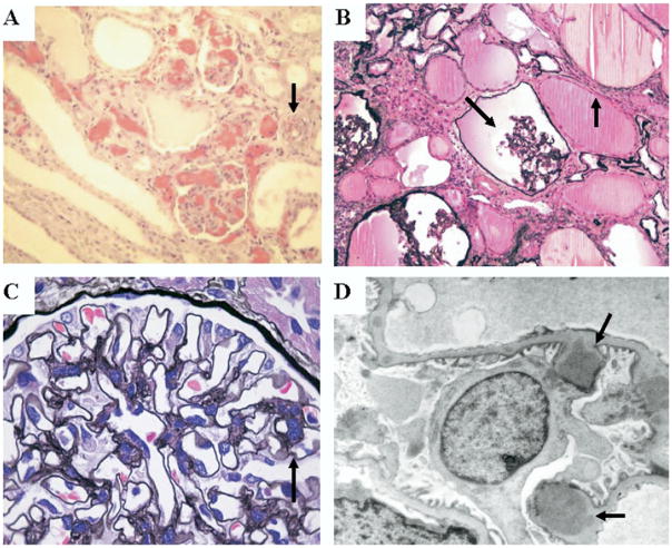
Figure 2.
Representative photomicrographs of renal sections from children with HIV-HUS, HIVAN, and HIVICK. (A) Light microscopy renal section from a child with HIV-HUS showing collapsed glomerular capillary loops with red blood cell fragments and tubular microcysts. The black arrow shows an arteriole with luminal narrowing owing to red cell fragments and intramural thrombosis (hematoxylin-eosin stain, 200×). (B) Light microscopy renal section from a child with HIVAN. The black arrows show a shrunken glomerulus and microcystic tubular dilatation (Jones methenamine silver stain, 200×). Figure 2B courtesy of Dr. William Bates and Dr. Peter Nourse. (C) Light microscopy renal section from a child with HIVICK showing mesangial prominence. The glomerular tuft is lobulated with double contours of the glomerular basement membrane (black arrow) (Jones methenamine silver stain, 700×). (D) Transmission electron microscopy from a glomerular capillary in a child with HIVICK. The black arrows show subepithelial deposits (4.000×). Figure 2C and D courtesy of Drs. Stewart Goetch and Professor Udai Kala.

