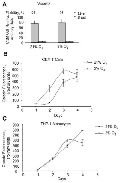
Fig. 3.
Effect of O2 concentration on cell viability and proliferation. A: Determination of CEM cells viability by trypan blue exclusion assay. CEM-GFP cells were grown in a 96-well plate, infected with Ad-Tat, and cultured at 21% O2 or physiological 3% O2 for 18 h. Cells were then supplemented with Trypan Blue and counted to determine cell viability by Trypan blue exclusion assay. The results are representative of three experiments. B and C: Determination of cellular proliferation by calcein-AM uptake assay. CEM cells (part B) or THP-1 cells (part C) were seeded in 96-well plates with threefold increment, cultured for the indicated amount of days at 21% O2 or 3% O2. The cells were supplemented with calcein-AM and calcein fluorescence was measured as described in Materials and Methods Section. The results are representative of three experiments.

