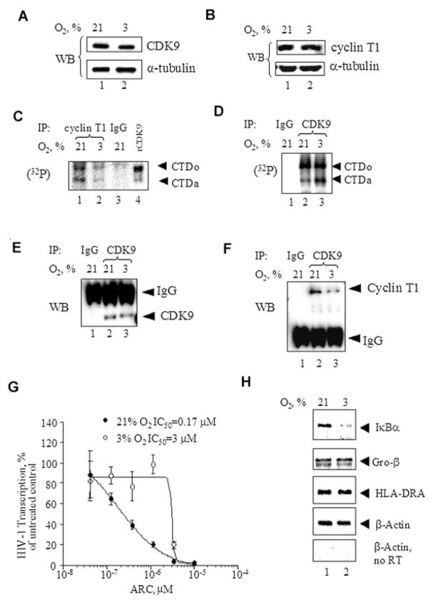
Fig. 7.
Effect of O2 concentration on cellular activity of CDK9. A and B: The effect of O2 concentration on the expression of CDK9 and cyclin T1. 293T cells were grown in 100 mm plates at 21% O2 (lane 1) or 3% O2 (lane 2) for 18 h. The equal amount of protein lysate was resolved on 10% SDS–PAGE and immunoblotted with the indicated antibodies. C: O2 concentration and CDK9/cyclin T1 activity. Cyclin T1 was immunoprecipitated from cell lysates prepared as in part A. The immunoprecipitated material was incubated with recombinant GST-CTD in the presence of γ-(32P) ATP. Kinase reactions were resolved on 10% SDS–PAGE and exposed to the Phosphor Imager screen. Lane 1, cells treated at 21% O2. Lane 2, cells treated at 3% O2. Lane3, preimmune IgGs were used for the immunoprecipitation. Lane 4, recombinant CDK9/cyclin T1 was used to phosphorylate GST-CTD. D: O2 and total CDK9 activity. CDK9 was immunoprecipitated from 293T cell lysates prepared as in part A. The immunoprecipitated material was incubated with recombinant GST-CTD in the presence of γ-(32P) ATP. Kinase reactions were resolved on 10% SDS–PAGE and exposed to the Phosphor Imager screen. Lane 1, preimmune IgGs were used for the immunoprecipitation. Lane 2, cells treated at 21% O2. Lane 3, cells treated at 3% O2. E and F: O2 concentration and dissociation of CDK9 and cyclin T1. CDK9 was immunoprecipitated from 293T cell lysates prepared as in part A. The immunoprecipitated material was resolved on10% SDS–PAGE and immunoblotted with antibodies against CDK9(part E) or cyclin T1 (part F). Lane 1, preimmune IgGs were used for the immunoprecipitation. Lane 2, cells treated at 21% O2. Lane 3, cells treated at 3% O2. All experiments are representative of at least three different experiments. G: O2 concentration and inhibition of HIV-1 transcription by ARC.293Tcells were grown in 96-well plates and transiently transfected with HIV-1LTR-LacZ vector with Tat-expressing vector (lane 2). The cells were treated with indicated concentrations of ARC and cultured at 21% O2 or 3% O2 for 18 h. Then the cells were lysed and analyzed for β-galactosidase activity. The results are triplicates and representative of two experiments. H: O2 concentration and expression of CDK9/cyclin T1-dependent genes. Semi-quantitative RT-PCR analysis of CDK9/cyclin T1-dependent genes. Total RNA was isolated from 293T cells cultured at 21% O2 and 3%, reverse transcribed and amplified with primers for IκBα, Gro-β and HLA-DRA and β-actin as described in Materials and Methods Section. PCR products were resolved on 2% agarose gel and visualized with ethidium bromide staining. No RT, control in which RNA was not reverse transcribed.

